p53 and Cell Cycle

In order for a living being to survive; their cells must go through a necessary process of replication and division.  This process is called the cell cycle.  The cell cycle is a multi-step cycle that includes the following phases: G1 phase, S phase, G2 phase, Mitosis, and cytokinesis.  Since the cell cycle is necessary for survival it must be tightly regulated.  If the cell cycle isn’t regulated properly, or if there are mutations in the DNA the cell can replicate and divide uncontrollably.  This uncontrollable cell grow will lead to cancer.  One of the key proteins that regulates the cell cycle is called p53.  The damaged or mutated DNA that will lead to the erratic cell grow is halted by p53.  The presence of p53 in the cell cycle of damaged DNA will lead to cell cycle arrest, DNA repair, or apoptosis.  To initiate the cell cycle arrest in response to damage DNA; p53 binds to two sites that are upstream of the p21 promoter, which will cause the creation of the p21 protein.  The p21 protein will eventually cause the cell cycle to arrest at the G1 phase by binding to the cyclin E/CDK2 complex.  The cell cycle arrest is induce to provide time for DNA repairs, if the damage is minor.  Another p53 induce gene, p53R2 is responsible for DNA repairs.  Induction of p53 cause many genes that are involved in the apoptotic pathway to become active.  Bnip3L, caspase6, Fas, Killer/Dr5, Puma, Noxa are just a few of the many genes p53 will induce to achieve apoptosis of a damage cell.  The importance of a check and balance system in the cell cycle can’t be appreciated enough; without it humans and other species will cease to exist.
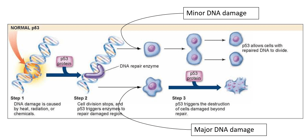
The image shows how p53 regulates the cell cycle when the DNA has minor and major damage.

Source:

https://schoolbag.info/biology/living/62.html

References:

  1. Chen, Jiandong. “The Cell-Cycle Arrest And Apoptotic Functions Of P53 In Tumor Initiation And Progression”. Cold Spring Harbor Perspectives In Medicine, vol 6, no. 3, 2016, p. a026104. Cold Spring Harbor Laboratory, doi:10.1101/cshperspect.a026104.
  2. “Primary Information Of P53 Gene”. Bioinformatics.Org, 2020, http://www.bioinformatics.org/p53/introduction.html.
  3. “The Cell Cycle, Mitosis And Meiosis — University Of Leicester”. Www2.Le.Ac.Uk, 2020, https://www2.le.ac.uk/projects/vgec/highereducation/topics/cellcycle-mitosis-meiosis.

p53 as Transcription Factor

The central dogma states that DNA is transcribed into RNA, which is then translated into proteins.  The central dogma must be under strict regulation, which is accomplished by transcription factors.  Transcription factors are molecules/proteins that control whether or not a gene is expressed (DNA made into protein).  As a transcription factor, p53 regulates the production/expression of many gene and gene products.  The expression of these products are induced by the activation of p53, which is a result of DNA damage.  There are many genes that are regulated by p53: bax, GADD45, MDM2, p21, Puma, and thrombosporin are all genes that are regulated by p53.  MDM2 is a gene that acts as a negative feedback mechanism for p53.  This negative feedback between p53 and MDM2 is to ensure that p53 isn’t overexpressed.  The cell cycle is also influenced by p53.  During the cell cycle there are many checkpoints to make sure that the DNA is replicated correctly.  If the DNA is damaged in anyway, p53 will be express, which will cause p53 to induce to production of either p21, GADD45, and Puma depending on the amount of damage done.  If minor damage, p21 will cause the cell cycle to stop so repairs can be made by the products of GADD45.  However, if the damage is too great to repair, Puma will initiate the apoptosis pathway.  This apoptotic pathway is a way to ensure that damaged/mutated DNA isn’t replicated.  Transcription factors are essential for the central dogma to continue, and for the survival of living beings.

The image depicts many gene that are regulated by p53; including the gene that is responsible for regulating p53 itself.

Source:

https://www.creativebiomart.net/researcharea-p53-pathway-proteins.htm

References:

  1. “Primary Information Of P53 Gene”. Bioinformatics.Org, 2020, http://www.bioinformatics.org/p53/introduction.html.
  2. Shaw, Phillip H. “The Role Of P53 In Cell Cycle Regulation”. Pathology – Research And Practice, vol 192, no. 7, 1996, pp. 669-675. Elsevier BV, doi:10.1016/s0344-0338(96)80088-4.
  3. “Gene Expression: What Are Transcription Factors?”. Thoughtco, 2020, https://www.thoughtco.com/what-are-transcription-factors-375675.
  4. Beckerman, R., and C. Prives. “Transcriptional Regulation By P53”. Cold Spring Harbor Perspectives In Biology, vol 2, no. 8, 2010, pp. a000935-a000935. Cold Spring Harbor Laboratory, doi:10.1101/cshperspect.a000935. Accessed 30 Jan 2020.

p53 and Cell Apoptosis

Once p53 has detected DNA damage due to mutations, it will decide whether the cell’s next step is apoptosis or cell repair.

Apoptosis, coordinated cell death, is one of the main controls for p53 tumor suppressor capability and increasing cell response to chemotherapy.

p53 can increase transcription of apoptotic genes such as Bcl-2 and BH3-only genes. The protein will bind to consensus sequences on these genes DNA segment and will induce Bcl-2 protein formation. Bcl-2 protein will then increase mitochondria apoptosis proteins.

p53 can increase caspase-6 protein production by binding to caspase-6 gene. Caspase-6 functions to increase cell reactivity to chemotherapy drugs.

p53 can increase sensitivity of cells to death receptor ligands and will thus encourage apoptosis directly and indirectly ( by means of “encouraging cell death in ligand rich environment” ) (Fridman)

p53 can be used as a gene response element, in which a minimal level of the protein will cause a protein to be induced.

ex. p-53 response gene, PIDD, is activated and will encourage cell apoptosis

p53 can increase ROS, reactive oxygen species, which will overall interrupt redox reactions within cell and cause mitochonidrial dysfunction to initiate cell apoptosis.

p53 can repress anti-apoptotic and mitotic genes

ex. Survivin, which as been implicated and over-expressed in many cancers

References

  1. Fridman, J., Lowe, S. Control of apoptosis by p53. Oncogene 22, 9030–9040 (2003). https://doi.org/10.1038/sj.onc.1207116

p53 and DNA Mutations

DNA damage can come in many forms: from wide-scale deletions of genes to the smaller instances of base-pair substitutions. UV and IR radiation are typical causes of disruptions in DNA strands.

p53 is a protein that is sensitive to all forms of DNA damage.

ex. p53 is activated with only a single break in double stranded DNA, which is typically caused by IR radiation.

” p53 contributes to genome maintenance to a large part by allowing time for the DNA-repair machinery to remove the lesions before cell proliferation resume”

Williams, Ashley B, and Björn Schumacher. “p53 in the DNA-Damage-Repair Process.” Cold Spring Harbor perspectives in medicine vol. 6,5 a026070. 2 May. 2016, doi:10.1101/cshperspect.a026070

p53 can by modified by reduction-oxidation couple reactions. Oxidative stress can induce DNA damage so it is crucial that p53 is consistently in a reductive form.

When there is cell with DNA damage: p53 will increase transcription of group of repair genes termed BER (base excision repair). These genes include DNA polymerase, OGG1, and AP endonuclease. Then p53 will modulate these BER proteins activity in a dose-dependent scheme of DNA damage. Low amounts of DNA damage will increase BER activity. High amounts of DNA damage will decrease BER activivty and p53-induced cell apoptosis will occur.

p53 binds to BER and other downstream target genes at their consensus sequence (10 base pairs). Phosphorylation of p53 at S315 and S378 will encourage DNA binding. Then the p53 tetramer will form allosterically when S392 is phosphorylated and S315 is dephosphorlyated.

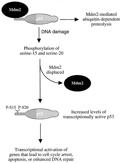
When there is cell without DNA damage: Another protein comes into play. Mdm2 is a regulator of p53. Mdm2 marks p53 for degradation by ubiquitin in the cytoplasm.

figure1

Source: Lakin, N., Jackson, S. Regulation of p53 in response to DNA damage. Oncogene 18, 7644–7655 (1999). https://doi.org/10.1038/sj.onc.1203015

References

  1. Lakin, N., Jackson, S. Regulation of p53 in response to DNA damage. Oncogene 18, 7644–7655 (1999). https://doi.org/10.1038/sj.onc.1203015
  2. Williams, Ashley B, and Björn Schumacher. “p53 in the DNA-Damage-Repair Process.” Cold Spring Harbor perspectives in medicine vol. 6,5 a026070. 2 May. 2016, doi:10.1101/cshperspect.a026070
  3. Yuangang Liu, Molly Kulesz-Martin, p53 protein at the hub of cellular DNA damage response pathways through sequence-specific and non-sequence-specific DNA binding, Carcinogenesis, Volume 22, Issue 6, June 2001, Pages 851–860, https://doi.org/10.1093/carcin/22.6.851

What is p53?

p53 is an extremely important protein in the cell cycle. This protein is known as the ‘master regulator‘ or the ‘guardian of the genome‘.

Image result for p53 protein"

Source: https://www.cgl.ucsf.edu/chimera/ImageGallery/entries/p53/p53.html

Image: p53 protein, with each of its four domains adherent to a DNA molecule, This multi-unit protein is comprised of over 300 amino acids in total.

This protein has multiple roles: recognize DNA mutations, act as a transcription factor for different genes, initiate cellular apoptosis, and control cell cycle.

Click links above to dive in to the world of cancer biology!

References:

  1. http://www.bioinformatics.org/p53/introduction.html

Create your own website, free on WordPress.com.Create your own website.
Start free